Watch What Happens to Your Brain When The Heart Pumps Blood
Your heart doesn’t exist in isolation, fiercely pumping blood while the rest of your viscera remain perfectly still. Each beat sends tiny shudders as blood moves around – and now a new MRI technique has managed to capture that effect on the brain in real-time, amplifying it to observe how it works.
It’s not just for funsies – although watching a video of the technique in action is absolutely fascinating – but to provide a tool that can help diagnose difficult-to-see conditions such as aneurysms and concussions.
The way this method helps diagnostics is by providing a baseline for the brain’s normal movement, which can therefore help doctors pick out anything unusual.
“We wanted to see if we could amplify the tiny movements of the brain with every heartbeat and capture that movement as it naturally occurs,” says mechanical engineer Mehmet Kurt of Stevens Institute of Technology in New Jersey.
“That’s important when you are trying to do what we are trying to do: detect abnormal motions in the brain to diagnose and monitor disorders.”
The amount by which your brain moves when your heart pumps blood into it is, in actuality, seriously minute: somewhere between just 10 and 150 micrometers, less than the width of the human hair. You wouldn’t want it to be a huge amount anyway, given the limited real estate in your skull.
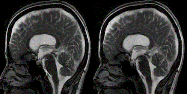
But this tiny motion is pretty hard to image with MRI. So the research team – comprising members from Stanford University, Stevens Institute of Technology, and the University of Auckland in New Zealand – found a way to magnify it.
They call their technique phase-based amplified MRI, and it has taken two years to refine. It works by combining a pulsometer, MRI and an algorithm developed at MIT for amplifying motion in real-world videos.
The patient wears the pulsometer, which allows the researchers to coordinate the heartbeat with images of the brain. The algorithm is then applied to the recording of the movement of blood and cerebrospinal fluid through the brain (the flow of the latter seems to correspond with the pulse).
Here’s where it gets really cool: the skull and all anatomical features are displayed at actual scale; the brain tissue motion, blood, and cerebrospinal fluid flow are magnified.
This gives doctors better visibility than the previous method of observing these changes, particularly in areas that move the most, such as the mid-brain and spinal cord. Using the new technique, these areas showed a significant reduction in video artefacts such as blooming and shading.
It also revealed subtle motion in other regions of the brain that had not been observed before.
To test their approach, the team tried it on two people: a healthy control, and a patient with a condition called Chiari malformation type I, whereby the brain tissue extends down into the spinal canal due to an abnormally shaped skull.
The brain of the Chiari patient showed significantly more movement in two locations compared to the healthy control.
The researchers plan to use phase-based amplified MRI in clinical settings on patients with a wide range of conditions, such as aneurysm, hydrocephalus, concussion, and structural brain abnormalities, to observe the different ways the brain moves in those cases.
“Better visualisation and understanding of the biomechanical properties of the brain could lead to earlier detection and monitoring of brain disorders,” Kurt said.
“It could also help with prevention, as it could lead to the design of better helmets for sports and recreation.”
Source:https://onlinelibrary.wiley.com/doi/10.1002/mrm.27236