Scientists Unlock Hidden Secrets of 2,300-Year-Old Mummies Using Cutting-Edge CT Scanner
High-tech imaging uncovered new insights into ancient mummies’ health and mummification methods.
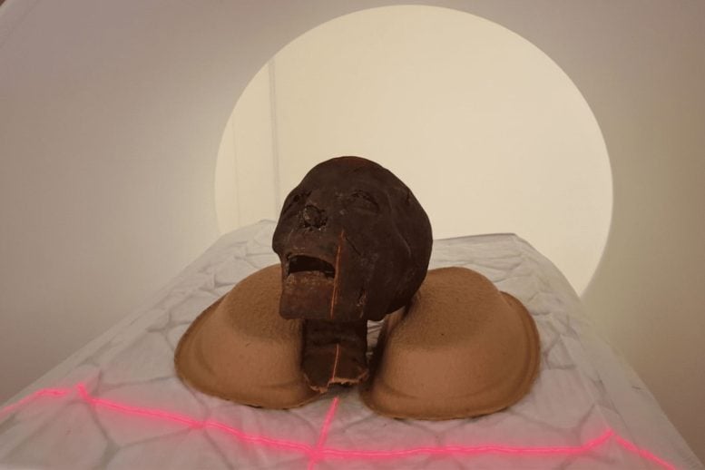
Egyptian mummy remains are being studied at Semmelweis University’s Medical Imaging Center (OKK) using one of the latest CT scanners with a photon-counting detector. The specimens, which come from the Semmelweis Museum of Medical History at the Hungarian National Museum Public Collection Centre (MNMKK), are producing highly detailed images that were not possible before, offering strong potential for new scientific discoveries.
Following standard clinical procedures, the scans were carried out at night outside regular patient hours. This advanced imaging method is especially effective for examining complex, layered materials and allows researchers to study mummified human remains in detail without causing damage.
“The aim of the examinations is to obtain as accurate a picture as possible of the internal structure of the remains, any abnormalities, and the preservation techniques used,” said Dr. Ibolyka Dudás, Chief Clinical Physician at the Department of Radiology and Head of the working group for post-mortem imaging.
Historical Context and Dating of the Remains
The Egyptian artifacts being analyzed have been part of the museum’s collection since its early years. Although they have undergone several imaging and multidisciplinary studies in the past, including conventional CT scans, earlier technology limited how much detail researchers could see. Radiocarbon (C14) dating was attempted on six samples, but only three produced usable results.
These results indicate that the oldest remains date from between 401 and 259 BCE, making them more than 2,300 years old. As part of the current project, all the museum’s Egyptian mummies, including this specimen, are being reexamined using improved imaging tools.
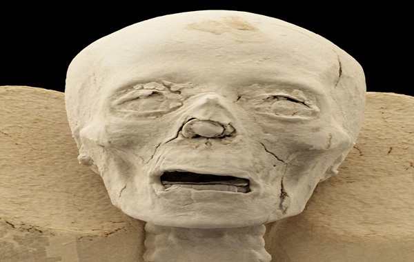
The new high-resolution scans provide a closer look at teeth and skull sutures in two mummified heads. This may help refine age estimates and support the creation of precise 3D models, including possible facial reconstructions.
New Medical and Biological Insights
For a left lower limb studied in the past, researchers had not been able to reach a clear diagnosis. The new images now suggest the individual may have had osteoporosis, although more analysis is needed to determine whether this was caused by aging or disease.
A second lower left limb appears to belong to a young person. While the exact age is still under investigation, this is the first time such detailed imaging has been available for this specimen.
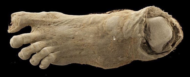
Another set of remains, once thought to be a simple mummy bundle, has produced surprising results. When it first entered the collection, it was identified as a human head and later possibly as a bird mummy. A previous CT scan clarified that it is actually an adult foot.
Mummification Techniques and Tissue Analysis
Researchers are also examining how textile fragments can reveal details about mummification practices, the individual’s age, and possible health conditions. The scans clearly show multiple layers of wrapping and their structural differences, which could support further historical and technical studies. The remains were likely once part of a complete mummy, though when and why they were separated is still unknown.
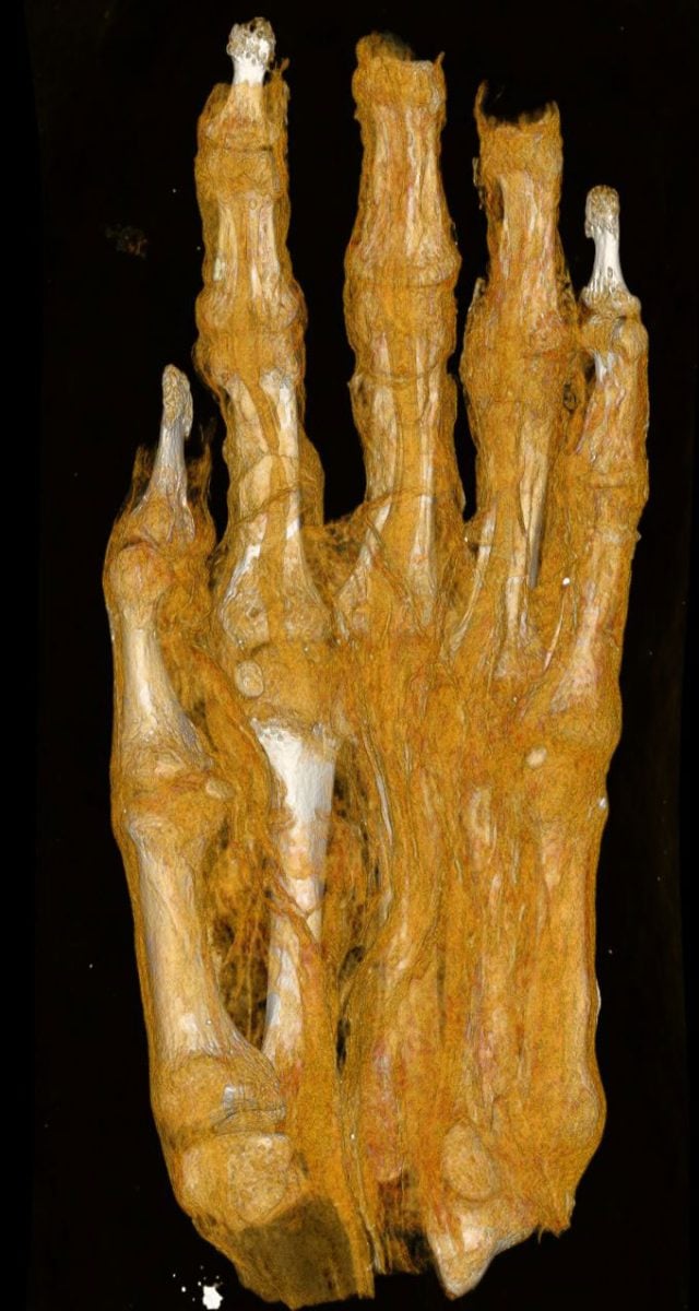
A mummified hand included in the study may also provide useful information. By analyzing bone size, development, and shape, scientists hope to determine whether it belonged to a child or an adult, as well as estimate sex and age.
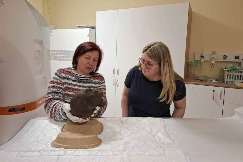
“The remains had previously been examined by a research team, but the current images provide a more detailed view than ever before and are expected to yield new, scientifically valid findings regarding the remains that have been preserved in the collection for decades,” emphasized the collection’s curator, Chief Museologist Krisztina Scheffer.
Future Research and Impact of Imaging Technology
Researchers are now carrying out a detailed review of the imaging data. They expect the analysis to shed new light on the mummies’ health, daily lives, and the techniques used to preserve them.
“Based on the results so far, it is evident that modern imaging technology opens up new perspectives in mummy research. It can reveal information hidden in finds that are thousands of years old without damaging them,” added the chief curator.
Source: science daily
42 lost pages of the new testament manuscript discovered
Scientists Unlock Hidden Secrets of 2,300-Year-Old Mummies Using Cutting-Edge CT Scanner