
Beyond Memory: The Brain’s Extra Fold Offers Key Insights into Frontotemporal Dementia
“Extra Fold” in the Human Brain May Delay Early Dementia for Years
Few people would have heard of frontotemporal dementia until earlier this year, when the family of actor Bruce Willis announced that the 68-year-old Bruce had been diagnosed with frontotemporal dementia.
Frontotemporal dementia is a rare disease, affecting only one in 20 people with dementia. Symptoms usually develop in the late 50s and first affect behavior, personality, and language skills. Unlike other dementias, memory loss occurs later in the disease.
People diagnosed with frontotemporal dementia usually die within eight years of diagnosis. Although the cause of frontotemporal dementia is largely unknown, approximately 30% of cases are hereditary. This also means that there are no treatments or cures to slow the progression of the disease.
However, a recent study I published with colleagues at Lund University may bring us one step closer to understanding the mechanisms underlying the onset and progression of frontotemporal dementia. We have discovered that the appearance of the brain may influence its resilience to the disease.
Folds of the Brain
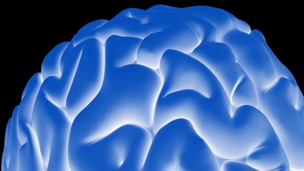
During pregnancy, the fetal brain forms characteristic folds as it grows in utero and expands within the skull. These brain folds play an important role in later cognitive functions.
The folds that form during early fetal development are found on both sides of the brain in all individuals. However, there are also folds that form later in development. This is called the parahippocampal sulcus and is not present in all individuals. This sulcus may be present on only one side of the brain, or it may be present on both sides.
The parahippocampal sulcus is an interesting one, the presence or absence of which causes significant differences in cognitive performance. For example, studies have shown that people who have the extraparietal sulcus on the left side and not on the right side have superior cognitive abilities.
A research team at Lund University, together with research teams in the United States and Amsterdam, has begun to study this role of the brain in dementia.
To really understand the role of the parafrontal sulcus, the research team decided to focus on a type of dementia in which brain damage occurs in the same region of the corpus callosum. The dementia chosen for this study is frontotemporal dementia. Frontotemporal dementia is an early-onset form of dementia that primarily affects the frontal lobes, particularly the central area around the cingulate sulcus.
Our team studied MRI brain images of 186 individuals diagnosed with frontotemporal dementia. Patients with frontotemporal dementia with a genetic cause were excluded. The results showed that approximately 57% of the subjects had a parahippocampal sulcus on the right side of the brain.
In subjects with this extra fold in the right side of the brain, symptoms of dementia were found to begin an average of two and a half years later. This may mean that the extraparietal sulcus delays the onset of symptoms. These results are statistically significant and indicate that they are not due to chance or other factors.
This 2.5 year delay in symptoms may not seem like much, but given the poor prognosis of the disease and the severity of its symptoms, it is a very meaningful time for patients and their relatives.
Cognitive Reserve
Nevertheless, patients with this extra cerebral lobe had an earlier onset of symptoms and a shorter survival time after symptom onset than those who did not. In other words, despite the delayed onset of symptoms, patients with and without this extra cerebral lobe died at similar ages.
While it may sound strange that one factor could delay symptoms while later hastening them, this paradox is an important feature of a principle known in neuroscience as “cerebral reserve.” Cerebral reserve is the structure of the brain that provides resilience to disease before symptoms appear. The critical point is the point at which the disease overcomes these defense mechanisms and the patient develops symptoms. After this critical point, people with higher brain reserve capacity deteriorate more rapidly than those with lower brain reserve capacity.
For example, people with higher education have higher brain reserve capacity and therefore develop Alzheimer’s disease later. Our study shows that the parahippocampal sulcus functions on a similar principle, first protecting people from symptoms and then progressing rapidly once symptoms begin.
Our study is the first to identify protective structures in the brain that delay the onset of symptoms in patients with frontotemporal dementia. Finding ways to maintain these protective structures could lead to the development of treatments to control symptoms and disease.
Source: Beyond Memory: The Brain’s Extra Fold Offers Key Insights into Frontotemporal Dementia
Beyond Memory: The Brain’s Extra Fold Offers Key Insights into Frontotemporal Dementia